|
Next to hand X-rays foot X-rays are an important investigation that patients with inflammatory arthritis. There is a need to have a systematic approach of reading a foot X-ray.
Normal foot X-rays
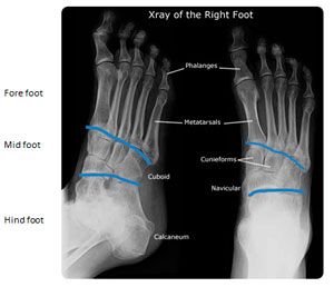
Usually AP and oblique views are taken.
 |
- X-ray feet AP and oblique
- AP: The fore foot and mid-foot are seen. The hind foot is not. The phalanges, metartarsal bones are seen
- Proximal interphalangeal joints, metatarso-phalangeal joints and mid-tarsal joints are seen.
- Hind foot bones and sub-talar joints are not seen.
After describing the type of X-ray one could proceed with talking about (1) bone health, (2) soft tissues, (3) joint space, (4) erosions and (5) malalignment |
Figure 1 |
|
 |
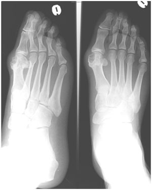
- X-Ray right foot AP and oblique
- Bone density is normal
- There is a soft tissue next to the first MTP join
- There is a reduction of joint space of the first MTP joint.
- There is a well-corticated cystic lesion/healed erosion at the head of the first metatarsal.
Diagnosis: Chronic gout |
Figure 2 |
|
 |
- X-ray right foot AP and oblique
- There is osteopenia
- Soft tissues are normal
- The DIP, PIP and MTP are well seen and normal
- There is a radio-dense lesion around a fracture in the second metatarsal bone
Diagnosis: Fracture with callus second metatarsal bone. |
Worksheet |
Fig. 1: |
|
 |
|
Fig. 2: |
|
 |
|
Fig. 3: |
|
 |
|
Fig. 4: |
|
 |
|
|
