|
previous answers |
Fig. 1: |
|
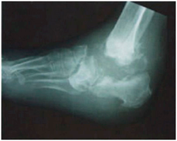 |
- X-ray of ankle in lateral view
- Complete destruction of talus. Significant destruction of the lower ends of tibia, fibula, and upper parts of calcaneus and proximal midtarsal bones.
- There is evidence of osteochondral fragments (arrow head).
DD 1. Infection in the ankle 2. Charcot joint |
Fig. 2: |
|
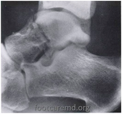 |
- X-ray of ankle in lateral view.
- There is a fracture in the neck of the talus.
|
Fig. 3: |
|
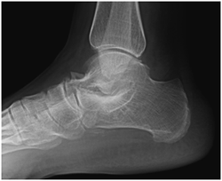 |
- X-ray of ankle in lateral view.
- Lower ends of the tibia and upper ends of talus are well seen.
- The subtalar joint is not well visualized.
- There is evidence of OA in the talonavicular joint with osteophyte formation.
Imp: Osteoarthritis talonavicular joint.
Subtalar joint not visualized because of flat feet.
|
Fig. 4: |
|
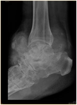 |
- X-ray of ankle in lateral view.
- Lower end of tibia is seen.
- The talus, talonavicular joint, upper part of calcaneus, and bones of the midtarsal joints are indistinct.
- There is a large area of soft tissue around the joint.
- There are osteochondral fragments in the soft-tissue.
Imp: Infection of the ankle. |
|
 |
|
 |
|