


Fellows’ corner

Dr Shefali Sharma, Rheumatology, PGIMER, Chandigarh
Hamman's crunch in Amyopathic Dermatomyositis
Dr Adarsh M B, Dr Preksha Dwivedi, Dr Varun Dhir, Dr Shefali K Sharma,
Dr A Sharma, Prof Sanjay Jain
Department of internal medicine, PGIMER, Chandigarh
23 year old male with symmetrical inflammatory polyarthritis and fever for last 2 years, presented with progressive breathlessness for last 3 months and acute worsening since last 5 days. On examination he had periorbital lilac rash and gottronssign(Fig 1,2). He had no muscle weakness. On auscultation Hamman’s crunch was audible and had subcutaneous emphysema in neck. HRCT of thorax showed pneumomediastinum, subcutaneous emphysema and organizing pneumonia in lower lobe(Fig 3,4). His Creatine kinase and LDH levels were normal and EMG was unremarkable. ANA was negative by IIF. Anti JO1, PM-Scl and U1RNP were not deteced by ELISA. He was given prednisolone 1 mg/kg and azathioprine. He improved clinically over next 1 month with disappearance of fever, rash, arthritis and the pneumomediastinum.
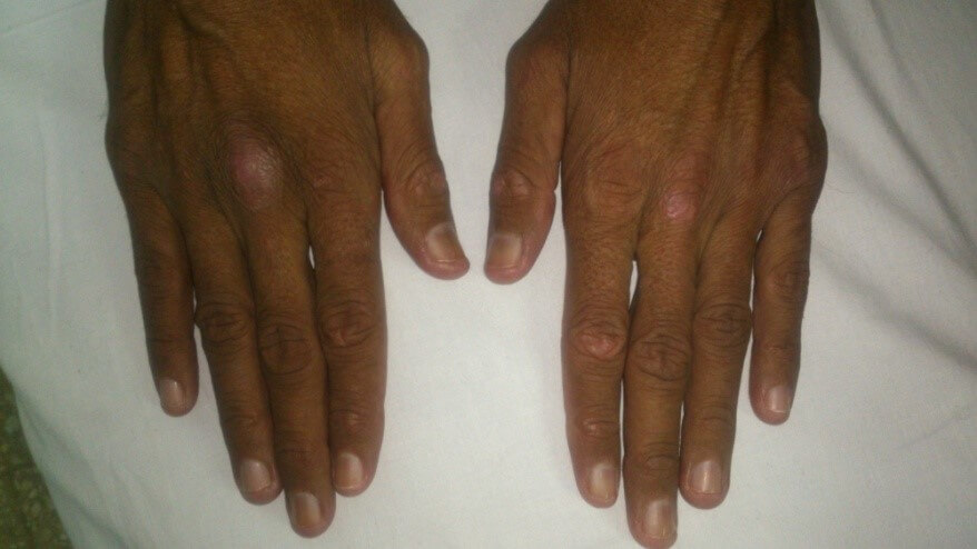
Fig 1 Gottron’s sign

Fig 2. Periorbital rash
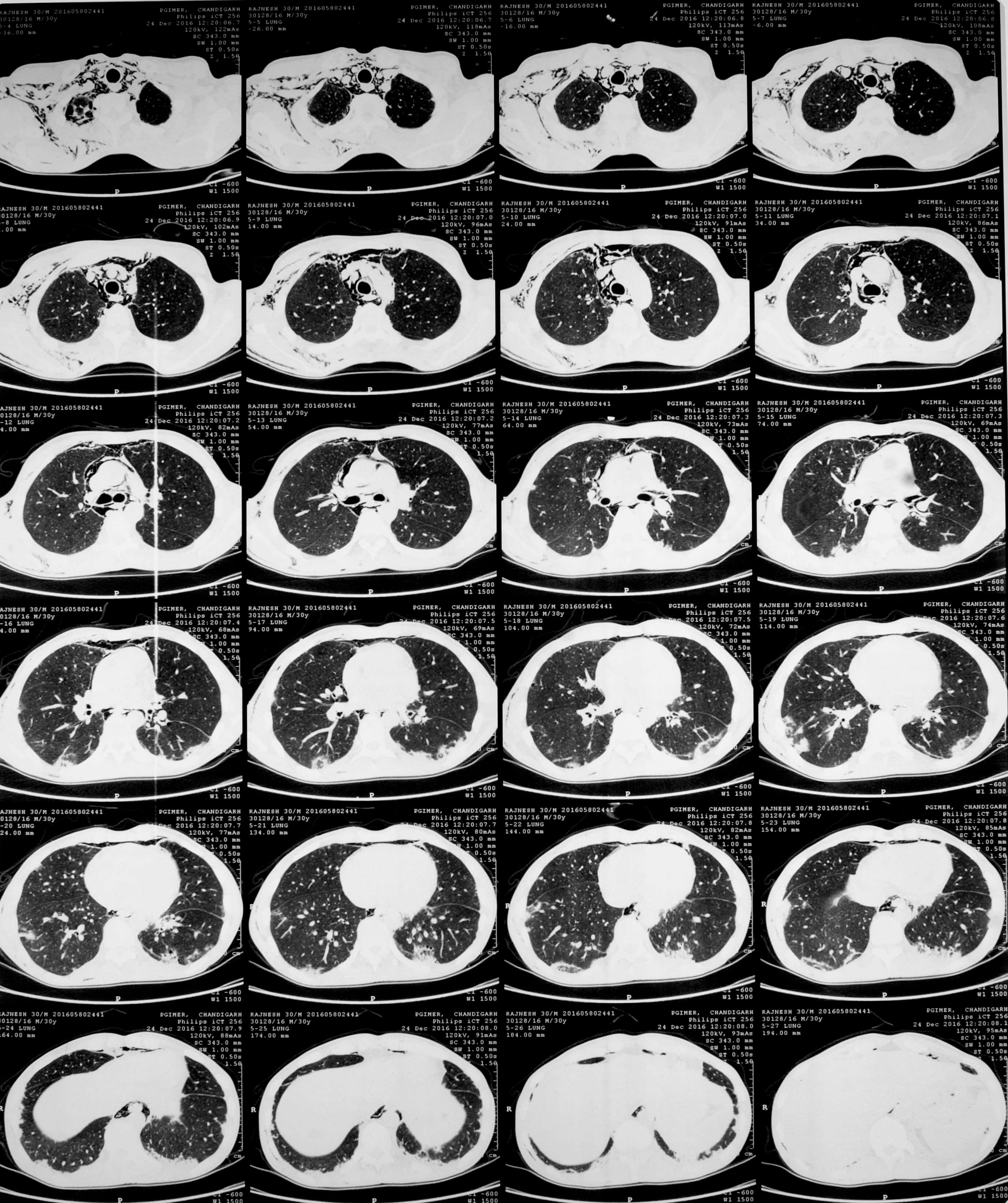

Fig 3,4. HRCT showing pneumomediastinum and subcutaneous emphysema
Recent studies in Chikungunya arthritis provide possible rationales for therapy
Dr Sanat Pathak
India has borne the brunt of 2 large chikungunya epidemics in the past decade, although literature about pathogenesis has been scarce. An estimated 25 – 50% have persistent joint pain after CHIKV infection. The recently published RHUMATOCHIK study followed 300 patients with rheumatologic manifestations for close to 3 years. Almost two-thirds of those infected had synovitisand joint pain persisted in more than 80% at 32 months. Most patients had functional improvement stressing the need for effective therapy in treating these patients.
Ravindran et al have previously shown the efficacy of combination DMARD (Methotrexate, Sulfasalazine and Hydroxychloroquine) in reducing both disease activity and disability. A randomized open label study is ongoing in PGIMER, chandigarh (NCT03058471) which will look at efficacy of starting Methotrexate early in Chikungunya Arthritis. Most trials in CHIKV arthritis have been empirical, considering the close similarity with RA.
Three studies published recently explore treatment options in a ‘bench to bedside’ manner. Using a CHIKV arthritis mouse model, Teo et al demonstrated the central role of CD4+ T cells in musculoskeletal manifestations. They went on to characterize the CD4+ T cells, and using a proteome wide screen, were able to identify two viral epitopes- nsP1 and E2 viral protein- that were recognized by these cells. This provided a rationale for drugs targeting T cells to be tested.
An unusual candidate- The S1P blocker fingolimod, approved for use in multiple sclerosis- proved to be effective, by reducing the migration of CD4+ T cells into the joint.
In another study, Miner et al tested 8 conventional DMARDs approved in rheumatoid arthritis in a similar mouse model, and found abatacept and the JAK inhibitor tofacitinib could curtail joint swelling and systemic inflammation. However the viral load in the joint persisted, and combination of Anti CHIKV monoclonal antibody and abatacept proved to be more efficacious in ameliorating the disease manifestations. This gives us indirect evidence of the importance of synovial persistence of virus in arthritis. The mouse model mirrors acute CHIKV arthritis in humans and the reproducibility of these studies in persistent arthritis is unknown, but they provide an impetus for further testing.
A group from NIV characterized NK populations in CHIKV arthritis and Rheumatoid arthritis. Patients with chronic arthritis showed higher expression of TNF-α producing NK cells while other NK cell subsets were reduced. Banking on the results of Anti TNF therapy in RA, this study may form the basis for the use of Anti-TNF agents in CHIKV arthritis as well.
Bouquillard E, Fianu A, Bangil M, Charlette N, Ribéra A, Michault A, Favier F, Simon F, Flipo RM. Rheumatic manifestations associated with Chikungunya virus infection: A study of 307 patients with 32-month follow-up (RHUMATOCHIK study). Joint Bone Spine. 2017 Feb 24. pii: S1297-319X(17)30034-9.
Teo TH, Chan YH, Lee WW, Lum FM, Amrun SN, Her Z, Rajarethinam R, Merits A, Rötzschke O, Rénia L, Ng LF. Fingolimod treatment abrogates chikungunya virus-induced arthralgia. Sci Transl Med. 2017 Feb 1;9(375).
Miner JJ, Cook LE, Hong JP, Smith AM, Richner JM, Shimak RM, Young AR, Monte K, Poddar S, Crowe JE Jr, Lenschow DJ, Diamond MS. Therapy with CTLA4-Ig and an antiviral monoclonal antibody controls chikungunya virus arthritis. Sci Transl Med. 2017 Feb 1;9(375).
Thanapati S, Ganu M, Giri P, Kulkarni S, Sharma M, Babar P, Ganu A, Tripathy AS. Impaired NK cell functionality and increased TNF-α production as biomarkers of chronic chikungunya arthritis and rheumatoid arthritis. Hum Immunol. 2017 Apr;78(4):370-374.
What was happening in rheumatology
70 years ago : The advent of measurement in rheumatology
In a field previously marked with protean symptoms and vague impressions, John Lansbury championed precision by his ideas about measurement of disease activity in rheumatoid arthritis. In the years between 1956 and 1958, he published a series of papers that laid the foundation of methods of quantification in RA that we use to this day. In the earlier papers, he conceptually outlined domains that could be used: rest pain, duration of early morning stiffness, weight, grip strength, Serum albumin and ESR among others. His major contribution comes from the development of the Lansbury index, the first articular index in RA. The process was laborious and cumbersome: he meticulously measured the surface area of each joint in a cadaver by using aluminium foil to cover cartilage. By weighing these pieces of foil, he came up with relative weights for each joint. Total inflammation could be estimated by the summation of individual values, each graded for the level of inflammation. In separate papers, he outlined a ‘systemic index’ and ‘motion index’ as well. The former was a composite score including anemia, ESR, Grip strength, Aspirin dose and fatigue. The motion index measured total lost motion as a percentage of possible motion, summated for all joints. Due to the tedium involved in applying them, none of these pioneering indices are in use today. However, they can rightly be considered forerunners of the numerous indices that have come in use in subsequent years, both in clinical trials and patient care- from the Ritchie articular index to simple joint counts to composite disease activity scores such as the CDAI. Importantly, Lansbury’s work – done prior to the advent of the widespread use of computers- gave us the paradigm that Rheumatoid arthritis can easily be quantified.
Lansbury J. Area of joint surfaces as an index to total joint inflammation and deformity. American Journal of the Medical Sciences 1956;232:150-155
Lansbury J. A method for summation of the systemic indices of rheumatoid activity.American Journal of the Medical Sciences 1956;232:300-310
50 years ago: The first successful candidate gene study in Rheumatoid arthritis
In the early 1970s, Stastny found that blood lymphocytes from RA patients showed a poor response against cells from other RA patients in mixed lymphocyte culture experiments, while they were responsive against lymphocytes from healthy controls. This suggested to him a shared histocompatibility determinant and he demonstrated that patients of RA shared an LD determinant named HLA-Dw4 (later named HLA DR4). The true validation of this came in a case control study where the candidate gene, HLADw4 was present in a much higher frequency in RA (59%) as compared to controls (16%). This was the first positive candidate gene study in RA. At a time when the immunologic nature of the disease was still debated, this association paved the way for further research of immune mediated mechanisms in general and T cell roles in particular, culminating in efficacious therapeutic options. The paper also highlighted the importance of ethnicity: the candidate gene was not present in higher frequencies in the few blacks included in the study.
Stastny P, Fink CW. HLA-Dw4 in adult and juvenile rheumatoid arthritis. Transplantation proceedings 1977;9(4):1863-1866

Dr Sanat Phatak, DM SGPGIMS, Lucknow
Fellows’ corner : From the bench
Dr Avinash Jain
Tregs restrain dendritic cell autophagy to ameliorate autoimmunity
Alissafi, Themis et al. "Tregs Restrain Dendritic Cell Autophagy To Ameliorate Autoimmunity". Journal of Clinical Investigation (2017): n. pag. Web. 25 June 2017.
Design of efficacious Treg-based therapies and establishment of clinical tolerance in autoimmune diseases have proven to be challenging. The clinical implementation of Treg immunotherapy has been hampered by various impediments related to the stability and isolation procedures of Tregs as well as the specific in vivo targets of Treg modalities. Herein, we have demonstrated that Foxp3+ Tregs potently suppress autoimmune responses in vivo through inhibition of the autophagic machinery in DCs in a cytotoxic T-lymphocyte–associated protein 4–dependent (CTLA4-dependent) manner. Autophagy-deficient DCs exhibited reduced immunogenic potential and failed to prime autoantigen-specific CD4+ T cells to mediate autoimmunity. Mechanistically, CTLA4 binding promoted activation of the PI3K/Akt/mTOR axis and FoxO1 nuclear exclusion in DCs, leading to decreased transcription of the autophagy component microtubule-associated protein 1 light chain 3β (Lc3b). Human DCs treated with CTLA4-Ig, a fusion protein composed of the Fc region of IgG1 and the extracellular domain of CTLA4 (also known as abatacept, marketed as Orencia), demonstrated reduced levels of autophagosome formation, while DCs from CTLA4-Ig–treated rheumatoid arthritis patients displayed diminished LC3B transcripts. Collectively, our data identify the canonical autophagy pathway in DCs as a molecular target of Foxp3+ Treg–mediated suppression that leads to amelioration of autoimmune responses. These findings may pave the way for the development of therapeutic protocols that exploit Tregs for the treatment of autoimmunity as well as diseases in which disturbed tolerance is a common denominator.
Ndfip1 restricts mTORC1 signalling and glycolysis in regulatory T cells to prevent autoinflammatory disease
AwoAkosuaKesewa Layman1,2,*, Guoping Deng3,*, Claire E. O’Leary3,*, Samuel Tadros3, Rajan M. Thomas4, Joseph M. Dybas4, Emily K. Moser3, Andrew D. Wells4, Nicolai M. Doliba5 & Paula M. Oliver3,4
Nature Communications June 2017
Foxp3 þ T regulatory (Treg) cells suppress immune cell activation and establish normal immune homeostasis. How Treg cells maintain their identity is not completely understood. Here we show that Ndfip1, a coactivator of Nedd4-family E3 ubiquitin ligases, is required for Treg cell stability and function. Ndfip1 deletion in Treg cells results in autoinflammatory disease. Ndfip1-deficient Treg cells are highly proliferative and are more likely to lose Foxp3 expression to become IL-4-producing TH2 effector cells. Proteomic analyses indicate altered metabolic signature of Ndfip1-deficient Treg cells and metabolic profiling reveals elevated glycolysis and increased mTORC1 signalling. Ndfip1 restricts Treg cell metabolism and IL-4 production via distinct mechanisms, as IL-4 deficiency does not prevent hyperproliferation or elevated mTORC1 signalling in Ndfip1-deficient Treg cells. Thus, Ndfip1 preserves Treg lineage stability and immune homeostasis by preventing the expansion of highly proliferative and metabolically active Treg cells and by preventing pathological secretion of IL-4 from Treg cells.
Protein arginine deiminase 4 inhibition is sufficient for the amelioration of collagen-induced arthritis
V C willis et al
Clin and Exp immunology May 2017
Citrullination of joint proteins by the protein arginine deiminase (PAD) family of enzymes is recognized increasingly as a key process in the pathogenesis of rheumatoid arthritis. This present study was undertaken to explore the efficacy of a novel PAD4-selective inhibitor, GSK199, in the murine collagen-induced arthritis model of rheumatoid arthritis. Mice were dosed daily from the time of collagen immunization with GSK199. Efficacy was assessed against a wide range of end-points, including clinical disease scores, joint histology and immunohistochemistry, serum and joint citrulline levels and quantification of synovial autoantibodies using a proteomic array containing joint peptides. Administration of GSK199 at 30 mg/kg led to significant effects on arthritis, assessed both by global clinical disease activity and by histological analyses of synovial inflammation, pannus formation and damage to cartilage and bone. In addition, significant decreases in complement C3 deposition in both synovium and cartilage were observed robustly with GSK199 at 10 mg/kg. Neither the total levels of citrulline measurable in joint and serum, nor levels of circulating collagen antibodies, were affected significantly by treatment with GSK199 at any dose level. In contrast, a subset of serum antibodies reactive against citrullinated and non-citrullinated joint peptides were reduced with GSK199 treatment. These data extend our previous demonstration of efficacy with the pan-PAD inhibitor Cl-amidine and demonstrate robustly that PAD4 inhibition alone is sufficient to block murine arthritis clinical and histopathological end-points.
Prevention of lupus nephritis development in NZB/NZW mice by selective blockade of CD28.
Laurent L1,2, Lefur A1,2,3, Bloas RL1,2, Néel M1,2, Mary C1,2,4, Moreau A5, Poirier N1,2,4, Vanhove B1,2,4, Fakhouri F1,2,3.
Eur J Immunol. 2017 Jun 20. doi: 10.1002/eji.201746923. [Epub ahead of print]
Abstract
Systemic Lupus Erythematosus (SLE) is a chronic systemic inflammatory disease. Autoantibodies (autoAbs) against double-stranded DNA (ds DNA), the hallmark of lupus, are produced and maintained by the interaction between auto-reactive B cells and CD4+ T cells. This interplay is controlled by the CD28/CD80-86/CTLA-4 axis. Here we investigated whether selective blockade of CD28-CD80/86 co-stimulatory interactions abrogates lupus nephritis development in a murine model of SLE. To this aim, NZB/NZW F1 mice were treated for 3 months, either with an anti-CD28 Fab' fragment or a control Fab'-IgG. The effect of CD28 blockade on lupus nephritis onset, survival, production of anti-ds DNA antibodies and costimulatory molecules was evaluated. CD28 blockade prevented the development of lupus nephritis and prolonged survival during the 3-month treatment and 12 weeks after. Furthermore, the production of anti-ds DNA autoAbs was decreased. Lastly, the protective effect of CD28 blockade was associated with increased intrarenal expression of the immunoregulatory molecule, Indoleamine 2, 3-dioxygenase, of the co-inhibitory receptor programmed cell-Death - 1 (PD-1) and of its ligand programmed death ligand - 1 (PDL-1).In conclusion, CD28 blockade prevented the development of lupus nephritis in NZB/NZW F1 mice. This immunomodulatory strategy is a promising candidate for SLE therapy in humans.

Dr Avinash Jain, DM resident, SGPGIMS, Lucknow