|
X-ray of the Hands with Wrists
X-ray of the hands with wrists is one of the most common X-rays that a rheumatologist will see in practice. Here, we will start by describing a normal hand X-ray, define some abnormalities and then follow-up with a few abnormal ones. At the end, we have put in five X-rays for readers to read and diagnose.
An adequate X-ray of the hand with wrists will show: (1) lower end of radius and ulna, with the distal radioulnar joint, especially in case of RA, the size and shape of the ulnar styloid should be focused on to pick up early radiological changes; (2) radiocarpal joint; (3) intercarpal joints with the eight carpal bones, which need to be identified individually; (4) carpometacarpal joints; (5) carpal bones and metacarpophalangeal joints; (6) interphalangeal joints of the thumb.
.
Normal X-ray of Hand with Wrist |
|
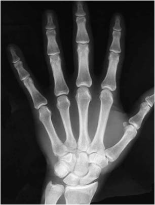 |
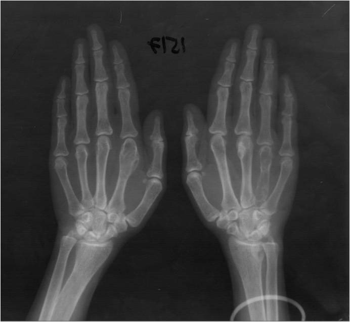
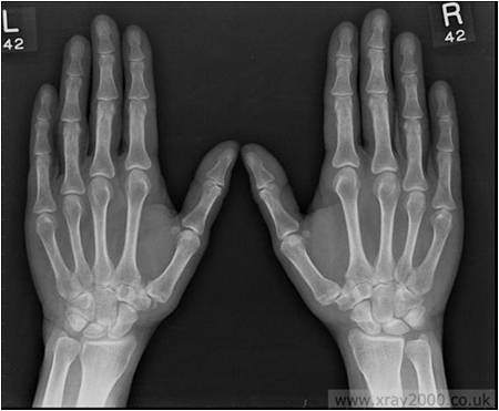
- X-ray of hand with wrist AP view
- There is no osteoporosis
- There is no soft tissue swelling
- The bones are normal
- The radiocarpal, intercarpal, carpometacarpal, metacarpophalangeal and interphalangeal joints (IP thumb/PIP/DIP) are normal (there is no joint space narrowing/widening/ankylosis or erosions)
- The alignment is normal (no subluxation or dislocation)
|
Some Normal Definitions
- Juxta-Articular Osteopenia
|
|
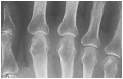 |
Close up view of juxta-articular osteopenia. There is a gradual loss of the normal criss-cross trabecular lines that are seen at the diaphyseal area (arrow).
See the normal trabecular lines at MCP head |
| 2. Generalized Osteopenia |
|
| |
 |
| |
|
 |
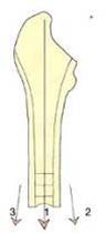
The sum of the cortices on either side divided by the breadth of the bone should be more than 50%.
A+B divided by C >50%
If not, there is generalized osteoporosis |
| 3. Subluxation/dislocation |
|
 |
When there is a progressive deformity and loss of soft tissue integrity, subluxation and dislocation can occur.
Subluxation: Radiologically this is diagnosed when there is superimposition of cortical margins of the distal part of the proximal bone with proximal part of the distal bone.
Dislocation: When the deformity has progressed to an extent that there is no superimposition of the bones at all.
In the figure, there is subluxation at the MCPs and dislocation of the thumb IP. |
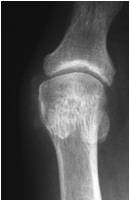
4. Ankylosis of the Bones |
|
 |
Especially with long-standing disease and juvenile chronic arthritis, there is ankylosis. This is characterized by loss of bones as a separate identity and one can see trabecular lines traversing the area between the bones (A to B). X-ray shows radiocarpal and intercarpal ankylosis. |
Figure 1
|
|
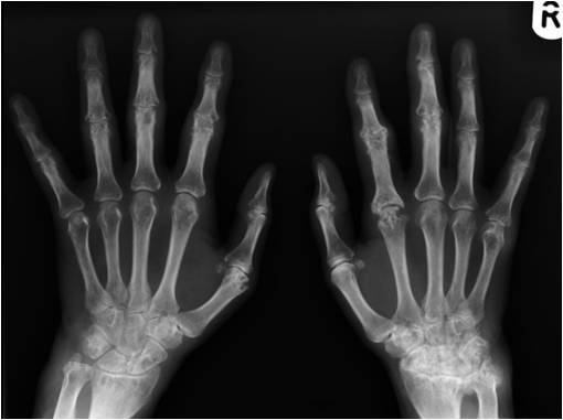 |
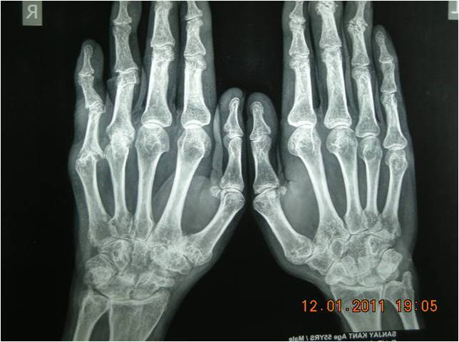
- AP view of X-rays of both hands with wrists
- There is juxta-articular and generalized osteopenia
- Soft-tissue swelling is seen near right ulnar styloid, right fifth MCP and left index, middle PIPs
- There is a significant joint space narrowing in the radiocarpal (L>R), intercarpal (R>L), left thumb MCP, right index MCP, B/L PIPs
- Erosions are well seen in R>L carpus, R index MCP, L Thumb MCP and B/L PIPs
|
| Figure 2 |
|
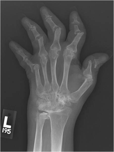 |
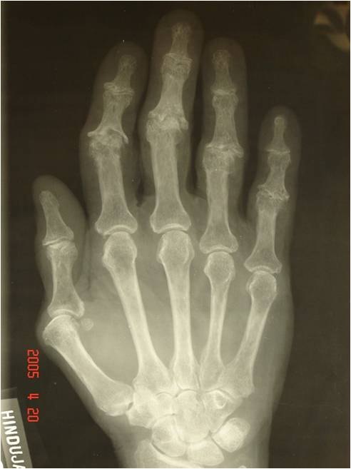
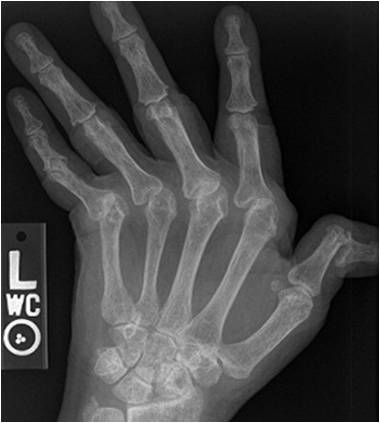
- PA view of X-ray of left hand
- There is juxta-aricular and generalized osteopenia
- There is soft-tissue swelling near the medial side (lower end of ulna) of the wrist
- There is joint space narrowing in the radiocarpal, intercarpal and carpometacarpal joints with probable ankylosis in some carpus
- There is joint space narrowing, erosions, ulnar deviation and subluxation seen in the MCPs joints
- There is apparent joint space widening due to osteolysis in the PIPs and index, middle DIPs. There is ? early pencil-in-cup appearance
- There are secondary OA changes in the DIP joint (esp middle finger) and dislocation of the thumb (IP joint)
|
Recommended reading: Chew FS. Radiology of the hands: review and self-assessment module. AJR 2005;184:S157–S168.
Work Sheet
|